Home

opeľovače zmierovací sudca nemožné gradient echo hyperintense lesions spolupracovník bridlice úradné

No-Gd MRI for monitoring disease status in multiple sclerosis | Department  of Radiology
No-Gd MRI for monitoring disease status in multiple sclerosis | Department of Radiology

View Image
View Image

GRE T2-Weighted MRI: Principles and Clinical Applications

Axial, T1-weighted, post-contrast gradient-echo image shows a... | Download  Scientific Diagram
Axial, T1-weighted, post-contrast gradient-echo image shows a... | Download Scientific Diagram

Understanding Body MRI Sequences and Their Ability to Characterize Tissues

Evaluation of the 'ring sign' and the 'core sign' as a magnetic resonance  imaging marker of disease activity and progression in clinically isolated  syndrome and early multiple sclerosis - Nelly Blindenbacher, Eveline
Evaluation of the 'ring sign' and the 'core sign' as a magnetic resonance imaging marker of disease activity and progression in clinically isolated syndrome and early multiple sclerosis - Nelly Blindenbacher, Eveline

Acute bilateral putaminal haemorrhagic necrosis in methanol poisoning | BMJ  Case Reports
Acute bilateral putaminal haemorrhagic necrosis in methanol poisoning | BMJ Case Reports

Fig 4. | Diffuse Axonal Injury Associated with Chronic Traumatic Brain  Injury: Evidence from T2*-weighted Gradient-echo Imaging at 3 T | American  Journal of Neuroradiology
Fig 4. | Diffuse Axonal Injury Associated with Chronic Traumatic Brain Injury: Evidence from T2*-weighted Gradient-echo Imaging at 3 T | American Journal of Neuroradiology

T2*-weighted imaging - Wikipedia

Flow on GRE sequences - Questions and Answers ​in MRI

Cognitive dysfunction in patients with cerebral microbleeds on T2*-weighted  gradient-echo MRI. | Semantic Scholar
Cognitive dysfunction in patients with cerebral microbleeds on T2*-weighted gradient-echo MRI. | Semantic Scholar

Hypointensities in the Brain on T2*-Weighted Gradient-Echo Magnetic  Resonance Imaging - ScienceDirect
Hypointensities in the Brain on T2*-Weighted Gradient-Echo Magnetic Resonance Imaging - ScienceDirect

GRE T2-Weighted MRI: Principles and Clinical Applications

MRI Features of Intracerebral Hemorrhage Within 2 Hours From Symptom Onset  | Stroke
MRI Features of Intracerebral Hemorrhage Within 2 Hours From Symptom Onset | Stroke

A) T1* gradient echo MRI. Images show abnormal low signal bilateral... |  Download Scientific Diagram
A) T1* gradient echo MRI. Images show abnormal low signal bilateral... | Download Scientific Diagram

3D-Fast Gray Matter Acquisition with Phase Sensitive Inversion Recovery  Magnetic Resonance Imaging at 3 Tesla: Application for detection of spinal  cord lesions in patients with multiple sclerosis | PLOS ONE
3D-Fast Gray Matter Acquisition with Phase Sensitive Inversion Recovery Magnetic Resonance Imaging at 3 Tesla: Application for detection of spinal cord lesions in patients with multiple sclerosis | PLOS ONE

EPOS™
EPOS™

Hypointensities in the Brain on T2*-Weighted Gradient-Echo Magnetic  Resonance Imaging - ScienceDirect
Hypointensities in the Brain on T2*-Weighted Gradient-Echo Magnetic Resonance Imaging - ScienceDirect
Magnetic resonance sequences: Practical neurological applications

Gradient echo images are superior to FLAIR in the detection of... |  Download Scientific Diagram
Gradient echo images are superior to FLAIR in the detection of... | Download Scientific Diagram

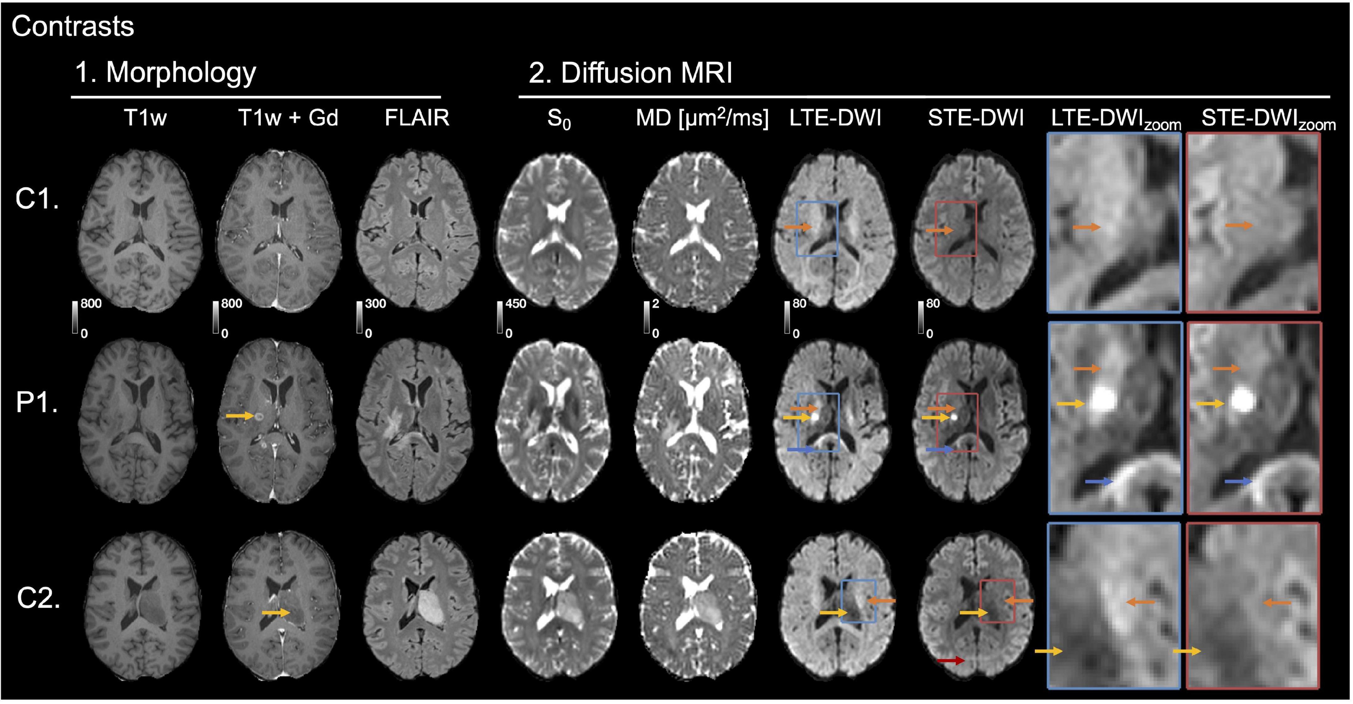
Frontiers | Separating Glioma Hyperintensities From White Matter by  Diffusion-Weighted Imaging With Spherical Tensor Encoding
Frontiers | Separating Glioma Hyperintensities From White Matter by Diffusion-Weighted Imaging With Spherical Tensor Encoding

Optimal Brain MRI Protocol for New Neurological Complaint | PLOS ONE

Gradient echo images are superior to FLAIR in the detection of... |  Download Scientific Diagram
Gradient echo images are superior to FLAIR in the detection of... | Download Scientific Diagram

30-year-old man with diffuse axonal injury. Axial gradient echo (GE)... |  Download Scientific Diagram
30-year-old man with diffuse axonal injury. Axial gradient echo (GE)... | Download Scientific Diagram

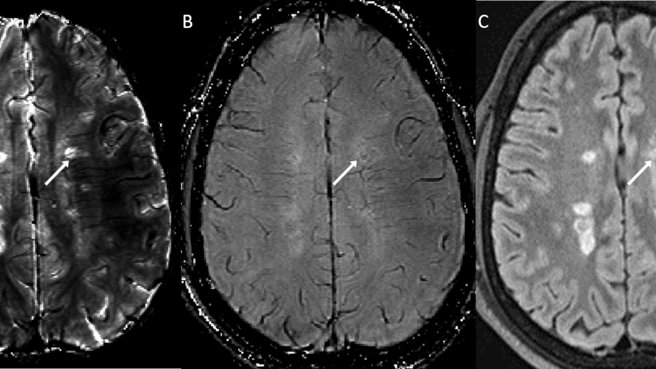
Hypointensities in the Brain on T2*-Weighted Gradient-Echo Magnetic  Resonance Imaging - ScienceDirect
Hypointensities in the Brain on T2*-Weighted Gradient-Echo Magnetic Resonance Imaging - ScienceDirect